Understanding the Melanocyte: Why Melasma Is More Than “Just Pigment”
Understanding the Melanocyte: Why Melasma Is More Than “Just Pigment”
One of the biggest shifts in my approach to melasma management happened when I stopped looking at melasma as a “pigment problem” and started looking at it as a melanocyte behavior problem.
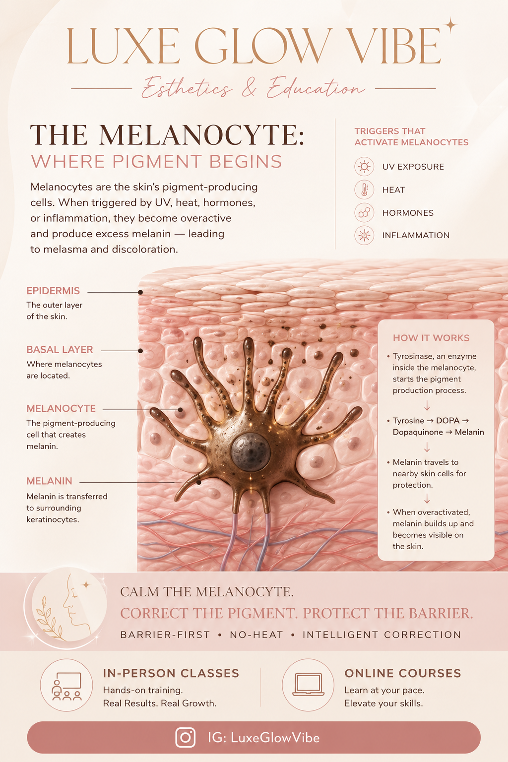
Melanocytes are the skin’s pigment-producing cells located in the basal layer of the epidermis. Their job is protective: they produce melanin and distribute it to neighboring keratinocytes through specialized structures called dendrites, forming what is known as the epidermal-melanin unit. Each melanocyte communicates with approximately 30–40 surrounding keratinocytes.
The challenge with melasma is that melanocytes become overreactive.These cells respond directly to UV exposure, heat, hormones, and inflammation through cellular receptors. When they perceive stress, they increase melanin production as a protective response. In melasma, this protective mechanism becomes dysregulated, causing pigment production to remain active longer than necessary.
Inside the melanocyte, the enzyme tyrosinase acts as the ignition switch for pigment formation:
Tyrosine → DOPA → Dopaquinone → Melanin
When this pathway remains activated by chronic inflammation, hormonal influences, heat exposure, or barrier dysfunction, excess pigment develops. Importantly, our goal is not to destroy melanocytes or “bleach” the skin, but to calm, regulate, and retrain melanocyte behavior.
Another concept I emphasize in corrective care is that melanocytes can develop a type of “memory.” Even after triggers are removed, chronic stimulation can cause melanocytes to continue producing pigment, which helps explain why melasma is often recurrent and requires long-term management rather than short-term correction.
A Quick Clinical Tip: Epidermal vs. Dermal Pigment
When assessing pigment visually under neutral lighting:
• Epidermal pigment typically appears brown and is located more superficially within the epidermis. It generally responds more favorably to topical correction and professional esthetic treatment.
• Dermal pigment often appears gray or gray-brown, reflecting pigment located deeper within the dermis. This presentation typically requires a slower, long-term management approach focused on calming inflammation, supporting barrier function, and regulating melanocyte activity.
For me, successful melasma management always comes back to the same principle:
Calm the melanocyte first. Correct the pigment gently. Protect the barrier always.
-Ivory S, LE